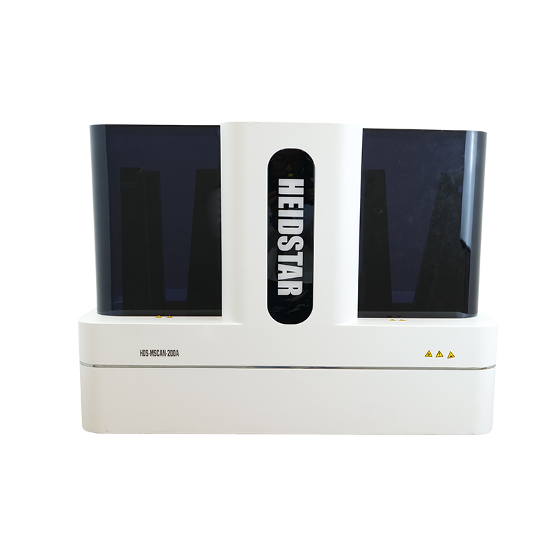
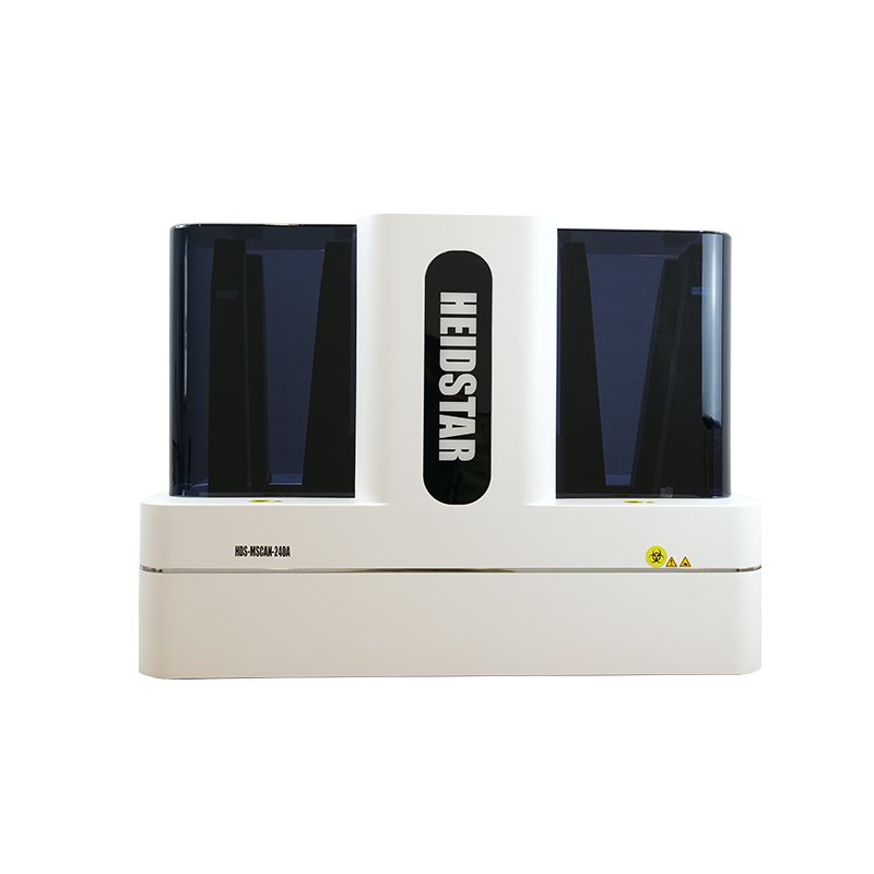
The Heidstar 4-slide digital pathology scanner is compact and portable, perfectly integrating various scenario applications, plug and play. It quickly scans large-area pathological sections, adopts advanced optical technology and intelligent image processing algorithms, ensuring high resolution of images and true color reduction, providing pathologists with clear and accurate diagnostic basis. In addition, digital slice images are easy to store and remotely transmitted, and support expert consultations at multiple locations, breaking geographical restrictions and improving the timeliness and accuracy of diagnosis. It also has OCR tag recognition technology, image stitching and Z-stack functions.
20000+ hours long-life LED light source, maintaining the same color temperature at all brightness levels.
Quickly browse the entire sample to improve observation efficiency.
With a high-precision sports platform, HDScanner scanning software can achieve one-click operation.
Kerke z-stack scan, seamless stitching and multi-layer fusion proofing to obtain high-definition images, supporting single-layer browsing.
As a specialized ODM partner in biomedical and precision optics fields, we deliver end-to-end customization services ranging from component-level design, mechanical structure optimization to automation control integration. We develop high-resolution fluorescence imaging systems and AI-powered pathology scanning platforms for global clients, offering comprehensive support from prototype validation to mass production.
Empower your brand with differentiated microscopy solutions that accelerate the journey from concept to clinical implementation.
Smart microscope suitable for daily work and research such as biomedical and veterinary medicine, microbiology, botany and forensic medicine.
Reliable and repetitive creation of digital slides, combining high-speed digital imaging and excellent image quality that conventional slide scanning systems cannot provide, as well as a wide variety of imaging modes.
It is used in research fields such as auxiliary drug discovery, cytotoxicity, cell tracking, tissue regeneration, cancer research, cell colocalization, etc.
Capacity | 4 |
Slides Dimensions | 25mmx75mm or 50mmx75mm or 110mmx75mm large slides |
Speed (Brightfield) | <40s (15mm*15mm, 20X objective) |
Scanning Camera | CMOS 5MP |
Preview Camera | 8MP |
Objective | 20x/ 0.75 NA (upgradeable to 40X) |
Pixel resolution | 0.25um/pixel(20X) |
Focusing Mode | Auto Focus |
Barcode Recognition | Supported |
Multi-layer Scanning | Supported |
Stage | Motorized XY Stage |
Image Format | JPEG/HDS/TIFF/BMP/SVS |
Brightfield Light Source | High-power long-life LED |
Dimensions (WDH) | 35cm x 39cm x 43cm |
Weight | 11kg |
Scanning Software | HDScanner multi-function scanning and recognition software (see software manual for details) |
Ready to bring your microscope imaging idea to life? Reach out to discuss your project or request a technical consultation.
ODM solution.