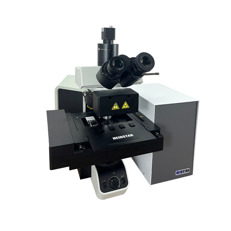
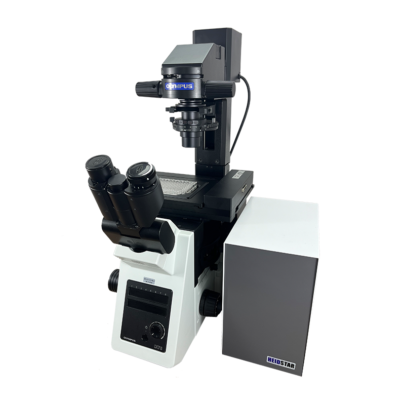
The Hydestar inverted fluorescence microscopy imaging scanning system is based on the OLYMPUS optical path, paired with a high-precision motion platform and an electric fluorescence turntable. Using HDScanner scanning software, it can realize one-click operation, automatic focus, automatic scanning, and collect low-noise, high-dynamic range specimens in real time, and can obtain sharp fluorescence images even under weak fluorescence.
It is equipped with a high-precision sports platform and electric fluorescent turntable, and uses HDScanner scanning software to achieve one-click operation.
As a specialized ODM partner in biomedical and precision optics fields, we deliver end-to-end customization services ranging from component-level design, mechanical structure optimization to automation control integration. We develop high-resolution fluorescence imaging systems and AI-powered pathology scanning platforms for global clients, offering comprehensive support from prototype validation to mass production.
Empower your brand with differentiated microscopy solutions that accelerate the journey from concept to clinical implementation.
Smart microscope suitable for daily work and research such as biomedical and veterinary medicine, microbiology, botany and forensic medicine.
Reliable and repetitive creation of digital slides, combining high-speed digital imaging and excellent image quality that conventional slide scanning systems cannot provide, as well as a wide variety of imaging modes.
It is used in research fields such as auxiliary drug discovery, cytotoxicity, cell tracking, tissue regeneration, cancer research, cell colocalization, etc.
Microscope | Olympus IX73 |
Slides Dimensions | 25mmx75mm or 50mmx75mm or 110mmx75mm large slides |
Speed (Brightfield) | 30mm/s |
Scanning Camera | Black & White CMOS 5MP |
Objective | Motorized, 20X0.5NA (see microscope configuration list) |
Pixel resolution | 0.16um/pixel (maximum resolution) |
Focusing Method | Manual / Auto (max focusing speed 200um/s) |
Barcode Recognition | Supported |
Multi-layer Scanning | Supported |
Stage | Motorized XY stage |
Image Format | JPEG/HDS/TIFF/BMP/SVS |
Repeating Positioning Accuracy | 2um |
Brightfield Light Source | LED |
Fluorescence Light Source | Full-band LED light source, integrated 4-channel fluorescence module, supports up to 8 channels, brightness adjustable via TTL signal |
Scanning Software | HDScanner multifunction scanning and recognition software (see software manual) |
Ready to bring your microscope imaging idea to life? Reach out to discuss your project or request a technical consultation.
ODM solution.